Fri, Apr 26, 2024
[Archive]
Volume 12, Issue 1 ( March 2020 2020)
Iranian Journal of Blood and Cancer 2020, 12(1): 29-33 |
Back to browse issues page

Download citation:
BibTeX | RIS | EndNote | Medlars | ProCite | Reference Manager | RefWorks
Send citation to:


BibTeX | RIS | EndNote | Medlars | ProCite | Reference Manager | RefWorks
Send citation to:
Fallah-Arzpeima S, Darbandi B, Hassanzadeh Rad A, Haghani Dogahe M, Niyasti P, Baghersalimi A. Multiple Cerebral Juvenile Xanthogranuloma; a Case Report. Iranian Journal of Blood and Cancer 2020; 12 (1) :29-33
URL: http://ijbc.ir/article-1-937-en.html
URL: http://ijbc.ir/article-1-937-en.html
Sima Fallah-Arzpeima1 
 , Bahram Darbandi2 , Afagh Hassanzadeh Rad3 , Mohamad Haghani Dogahe4 , Parham Niyasti4 , Adel Baghersalimi * 5
, Bahram Darbandi2 , Afagh Hassanzadeh Rad3 , Mohamad Haghani Dogahe4 , Parham Niyasti4 , Adel Baghersalimi * 5
1- Assistant Professor of Radiology, Department of Radiology, School of Medicine, Alzahra Hospital, Poursina Hospital, Guilan University of Medical Sciences, Rasht, Iran
2- Associated Professor of pediatric hematology and oncology, Pediatric Diseases Research Center, Guilan University of Medical Sciences, Rasht, Iran
3- PhD of Linguistics, Pediatric Diseases Research Center, Guilan University of Medical Sciences, Rasht, Iran
4- Student of Medicine, Guilan University of Medical sciences, Rasht, Iran
5- Assistant Professor of pediatric hematology and oncology, Pediatric Diseases Research Center, Guilan University of Medical Sciences, Rasht, Iran , bahersalimi498@yahoo.com
Abstract: (3112 Views)
Juvenile xanthogranuloma (JXG), is the most common form of non-Langerhans cell histiocytosis. It is a rare and usually benign, monoclonal proliferative disorder of histiocytic cells. The occurrence of multiple brain lesions is extremely rare. Due to the rarity of CNS disease, the preferred treatment and overall prognosis of these patients remain unclear. In this case report, authors present their experience about a patient with isolated multiple cerebral JXG which yields considerable outcomes. The patient was an eleven-year-old boy with anorexia and recurrent vomiting. Brain MRI demonstrated multiple lesions involving right lateral ventricle wall and periventricular white matter, the right side of splenium, mammillary body and tuber cinereum, fourth ventricle floor and roof, and cerebellar tonsil. After initial response to chemotherapy, the lesions recurred and radiotherapy was done. Consequent to good response after radiotherapy and despite the occurrence of a seizure attack, long term follow-up showed the marked resolution of the lesions and a good general status.
: Case report |
Subject:
Pediatric Hematology & Oncology
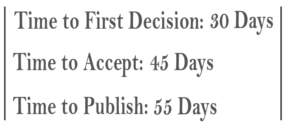
Received: 2019/09/17 | Accepted: 2020/04/19 | Published: 2020/05/2
Received: 2019/09/17 | Accepted: 2020/04/19 | Published: 2020/05/2
Send email to the article author
| Rights and permissions | |
 |
This work is licensed under a Creative Commons Attribution-NonCommercial 4.0 International License. |
- Iranian Journal of Blood and Cancer
- Journal Tel: +989369936116
- Publisher Tel: +982122275917
- Mail: Info@ijbc.ir