Mon, Nov 17, 2025
[Archive]
Volume 16, Issue 1 (March 2024 2024)
Iranian Journal of Blood and Cancer 2024, 16(1): 78-88 |
Back to browse issues page


Download citation:
BibTeX | RIS | EndNote | Medlars | ProCite | Reference Manager | RefWorks
Send citation to:


BibTeX | RIS | EndNote | Medlars | ProCite | Reference Manager | RefWorks
Send citation to:
Mardani Valandani H, zahedi A M, Mirzaee Khalilabadi R. The Influence of Extracellular Vesicles from Human Peripheral Blood Mononuclear Cells and Umbilical Cord Mesenchymal Stem Cells on Acute Lymphoid Leukemia Cells. Iranian Journal of Blood and Cancer 2024; 16 (1) :78-88
URL: http://ijbc.ir/article-1-1369-en.html
URL: http://ijbc.ir/article-1-1369-en.html


1- Department of Hematology and Medical Laboratory Sciences, Faculty of Allied Medical Sciences Kerman University of Medical Sciences, Kerman, Iran
2- Department of Hematology and Medical Laboratory Sciences, Faculty of Allied Medical Sciences Kerman University of Medical Sciences, Kerman, Iran ,Khalilabadi60@gmail.com
2- Department of Hematology and Medical Laboratory Sciences, Faculty of Allied Medical Sciences Kerman University of Medical Sciences, Kerman, Iran ,
Abstract: (1477 Views)
Objective: Many studies have suggested Mesenchymal stem cells, as a promising way to develop new treatment strategies for different types of disease. However, due to their possible tumorigenic effects, their clinical use has been limited. Since a great deal of MSCs’ therapeutic benefits depend on MSC-derived extracellular vesicles, these particles have been receiving much attention in the past couple of years. With this in mind, we aimed to study the Effects of both peripheral blood mononuclear cells and human umbilical cord MSC-derived extracellular vesicles on cell growth, proliferation, and apoptosis of the Nalm6 cell line.
Materials and Methods: Isolated HUCMSCs were cultured, and PBMCs were acquired using the Ficoll-Hypaque technique. Their EVs were then extracted. The Nalm6 cells were divided into five groups: a control group and four treatment groups, which were treated with MSC- and MNC-derived EVs at different concentrations. Cell viability and metabolic assays were evaluated using trypan blue staining and the MTT assay, respectively. Thereafter, a flow cytometric assay was performed to detect cell cycle progression and apoptosis.
Results: Our research revealed that the level of metabolic activity between Nalm6-treated EVs and the control group was not significantly different after 3 days. Also, no significant changes were reported in the growth and apoptosis effects of EVs on Nalm6 cells compared to the control group. Furthermore, different concentrations of EVs didn’t cause any inhibition on tumor growth.
Conclusion:Obtained EVs neither showed any anti-tumor effect nor caused any progression or aggressiveness in the Nalm6 cell line.
Keywords: Mesenchymal Stem Cells, Extracellular Vesicles, Umbilical Cord, Cell Survival, Cell Cycle.
: Original Article |
Subject:
Adults Hematology & Oncology
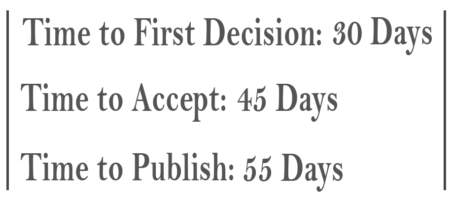
Received: 2023/11/15 | Accepted: 2024/01/20 | Published: 2024/03/25
Received: 2023/11/15 | Accepted: 2024/01/20 | Published: 2024/03/25
Send email to the article author
| Rights and permissions | |
 |
This work is licensed under a Creative Commons Attribution-NonCommercial 4.0 International License. |
- Iranian Journal of Blood and Cancer
- Journal Tel: +989369936116
- Publisher Tel: +982122275917
- Mail: Info@ijbc.ir